Catatan

Tunjukkan catatan dari 2017

E-Portfolio: Microbiology - Week 14

Imej
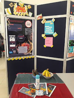
Assalam and hi. So, this week is our last week for this semester. We learned about Microbial Growth, Microbial Metabolism and the Nutritional Requirement of the microbes. This week also, we had our 3 Minute Talk (3MT) session for our Adopt A Microbe programme. Basically, everyone need to talk about what they know or they learn or they understand on their chosen microorganisms, For me, I chose Paenibacillus vortex. P. vortex  is actually a very useful bacteria especially for plants. They are a part of rhizosphere community that live with the plant and help fighting the plant pathogens. They have wide range of antimicrobial properties that can use to make antibiotics in order to kill other species of bacteria. So, here is my poster/slide of the talk.   At the end of our class, we had a small feast as Dr Wan bought us cakes and beverages. The chocolate cake was so goooooood! We took pictures together with Dr Wan. It was a good ending for this semester. Thank you all!

E-Portfolio: Microbiology - Week 13

Imej
Hello all! This week is such a hectic week to begin with. It was really tiring but it really worth and I'm so thankful to be a part of this week. For this week, we learned about Disinfectant and Sanitation, we also learn on Microbial Growth. But the highlight of this week is the Microbes Invention, Innovation and Ideation Challenge where the courses that took part in this program are Microbiology students, Biotechnology students and the Biocellular students. We teamed up to came up various products that use microorganisms for the production. For my team, we chose Chitosan Soluble Pills where we extract chitosan from chitin of the fungus, Rhizopus oryzae. This product is actually used to lengthen the shelf life of agricultural products especially fruits that undergoes ripening in short period of time. The chitosan form a coating for the fruits that lengthen their ripening period and actually consumable. It is also biodegradable and nature-friendly. Here is some photos from our MI

E-Portfolio: Microbiology - Week 12

Imej
Hi peeps! This week, Dr Wan taught us more about Viruses, Viriods and Prions. Basically, a virus is an infectious particle that reproduces by commandeering a host cell and using its machinery to make more viruses. A virus is made up of a DNA or RNA genome inside a protein shell called a capsid. Some viruses have an internal or external membrane envelope. Viroid is a piece of RNA without a protein coat responsible for several plant disease. Prions is consist of single type of protein with no nucleic acid component.  We also were given a group task to find information on a single virus of our choice. My group chose the Rabies virus.  Rabies virus is a rod- or bullet-shaped, single-stranded, negative-sense, unsegmented, enveloped RNA virus. The virus genome encodes five proteins.  Placement within the family is based on the distinctive morphology of the virus particle. Cross- reactive nucleoprotein antigens or comparative genomic sequences determine inclusion in the genus  Lyssavir

E-Portfolio: Microbiology - Week 11

Imej
This week instead of having class in the lecture room, we had our first road trip!!  We went to Institut Biosains (IBS), a place where the post graduate students done their researches and what not. Basically, the main reason we went to IBS is to learn more on Electron microscopy. Mr. Nazmi, a master degree student was the one who leads and guide us during the trip.  There, we met Kak Farah who is one of the staffs at IBS. She explained to us one the Scanning Electron Microscope, the Transmission Electron Microscope, the Ultramicrotome process and also the processes needed to be done for the sample to be observe under the microscope. She is such a friendly and nice person.  Here are some picture I want to share with you guys.  During our lab session this week, we had done the experiments on oxygen requirement for microbial growth and anaerobic system.  Basically, there are aerobic and anaerobic microbes that can be classified into:  Aerobic: Aerob

E-Portfolio: Microbiology - Week 10

Imej
Rise and shine, people! This week, we learned on Kingdom Protists that consist of: 1. Protozoa - consists of four main group which are Archaezoa, Rhizopoda, Ciliophora and Apicomplexa.  These  organism, usually single-celled and heterotrophic (using organic carbon as a source of energy), belonging to any of the major lineages of protist  and, like most protists, typically microscopic. All protozoans are eukaryotes  and therefore possess a “true,” or membrane-bound, nucleus 2. Algae - there are six groups of algae which are Chlorophyta (green algae), Rhodophyta (red algae), Phaeophyta (kelps), Pyrrophyta (dinoflagellates), Crystophyta (diatoms) and Euglenophyta. Algaes can be classified with several characteristics including their reproductive structure, chlorophyll and pigmentations, their storage materials, types of cell wall and others. 3. Slime molds -   are a type of protist that aggregates into colonies and ingest bacteria, fungal spores, and possibly other protists.  

E-Portfolio: Microbiology - Week 9

Imej
Assalam and hi! Here is the new post for this week.  This week, we learned on Fungi, one of the eukaryotes that has its kingdom. On Wednesday, our group had a discussion with our seniors who were taking Mycology class this semester. Our group discussed on Phaffia rhodozyma. Phaffia rhodozyma is a fungi from Basidiomycota group that is known for its astaxanthin. Astaxanthin is widely used in aquaculture especially to enhance the red pigment of salmon and trouts.  During our lab session this week, we had done our practical on nutritional requirements of several species of microbes. We used different type of broth and agar plate to culture the microorganisms. The results are quite fascinating as you see the same species of microorganism can produce beautiful colony on different cultures.  Different type of agar plates with different species of microorganisms.  E. coli colony with green sheen pigmentation on Eosin-methylene blue agar plate. 

E-Portfolio: Microbiology - Week 8

Imej
November 12, 2017  During our class with Dr. Wan this week, we learned through quizzes and games made by every group. The topic we emphasizes on is still on the internal structure of microorganisms. Here is the link for the quiz frommy group. Do try it to test you knowledge on this topic :) https://play.kahoot.it/#/k/5a939211-5475-4872-9dd4-698e3790d0fb  On Friday, we had done discussion in groups also regarding this topic. Our group had done about Golgi apparatus. We had given a task to look for any journal regarding the organelle we pick and make a summary from the journal. This is the link to the journal :  http://sci-hub.cc/http://www.jbc.org/content/270/9/4612.short   And here is the summary of the journal from our group.  "Giardia lambia  is a parasitic protozoan that cause Giardiasis, more commenly known as beaver fever with symptoms such as diarrhea, abdominal pain and weight loss. It spreads through cyst which will later germinate into tropozoite. This pro

E-Portfolio: Microbiology - Week 7

Imej
What is up, people! This week, we learned on internal structure and its function of a prokaryote with Dr. Wan Courtesy: https://www.shmoop.com/biology-cells/prokaryotic-cells.html I am actually quiet interested with plasmid, a small structure, circular, double-stranded DNA molecule that is distinct from a cell's chromosomal DNA. Plasmids naturally exist in bacterial cells, and they also occur in some eukaryotes. Often, the genes carried in plasmids provide bacteria with genetic advantages, such as antibiotic resistance. Plasmids have a wide range of lengths, from roughly one thousand DNA base pairs to hundreds of thousands of base pairs. When a bacterium divides, all of the plasmids contained within the cell are copied such that each daughter cell receives a copy of each plasmid. Bacteria can also transfer plasmids to one another through a process called conjugation using sex pili. For our experiment this week, we had done Gram staining and Acid-fast staining.

E-Portfolio BMY3101 - Week 6

Greetings! So for this week we learned on the external structures of prokaryotes. Basically there are several parts of external structures in prokaryotes which are : 1. Glycocalyx - h ighly-hydrated fibrous meshwork of carbohydrates that projects out and covers the membrane of endothelial cells to  mediatescell attachment, retains humidity during exposure to dry environments, protects against molecular and cellular antibacterial agents (antibiotics, surfactants, bacteriophages, phagocytes) and other vital functions 2. Flagella -    long, hairlike organelles that extend from the cell, permitting it to move. In prokaryotic cells, such as bacteria, the flagella rotate like the propeller of a motorboat 3. Axial filaments -  a filament that wraps around the cell and makes the cell move in a cork screw movement. It  composed of endoflagella that spiral around the cells instead of towards the surrounding meduim 4. Cell wall -  Bacteria are divided into two major groups:

E-Portfolio BMY3201 - Experiment 7

Imej
Hello awesome people! This week we played with colours during our experiment. I love it so much! Firstly, we learned on how to prepare smear of various cultures on the glass slides. Then, we did the steps for Simple staining and Negative staining.  Simple staining is a very simple staining procedure involving only one stain used to determine cell shape, size, and arrangement. Basic stains, such as methylene blue, carbol fuschin, or crystal violet are useful for staining most bacteria.  The negative stain is particularly useful for determining cell size and arrangement. It can also be used to stain cells that are too delicate to be heat-fixed. Nigrosin is used in this technique is an acidic stain.   Since the surface of most bacterial cells is negatively charged, the cell surface repels the stain. The glass of the slide will stain, but the bacterial cells will not. The bacteria will show up as clear spots against a dark background. Here is the observation of the stainin

E-Portfolio BMY3101 - Week 5

Imej
This week we were given a group task to choose a prokaryote and to make a presentation about the prokaryote. We had chose Serratia marcescens which is  a motile,short rod-shaped, Gram-negative, facultative anaerobe bacterium, classified as an opportunistic pathogen. It was discovered in 1819 by Bartolomeo Bizio in Padua, Italy. Bizio named the genus Serratia in honor of and Italian physicist named Serratia, and chose marcescens for the species name after the Latin word for decay.   S. marcescens was first thought to be harmless (non-pathogenic). Due to its ability to produce red pigmentation, it was first used in 1906 as a marker in order to trace bacterial activity or transmission. It was not until later in the 1950’s that the US government experimented with the S. marcescens and the harmful effects that the bacteria causes were revealed. A study using S. marcescens was carried out to determine the possibility of biological weapons being transmitted by wind current.   S. m

E-Portfolio BMY3201 - Experiment 6

Imej
11th October 2017 For Experiment 6, we learned on how to measure microorganism under the microscope. Basically we used an ocular micrometer, a lens with scale from 0 to 100 and stage micrometer, a slide with scale of 0.01 micrometer. The ocular micrometer was put inside one of the ocular lenses before observing. Ii get me took quite some times before I figured out on how to measure the microbes. We measured Staphylococcus aureus , Bacillus cereus and Serratia marcescens. Here are some pictures of the microbes under the oil immersion lens. This is Serratia marcescens under oil immersion lens This is  Staphylococcus aureus  under oil immersion lens

E-Portfolio BMY3101 - Week 4

Imej
Assalam and hi :) This week, Dr Wan taught us a little bit about Scanning Electron Microscope (SEM) and Transmitting Electron Microscope (TEM). Those microscope are something that were really expensive and only professionals can handle them. She promised us on wanting to take us on a field trip to see the SEM here in the UPM. I am so excited for the trip! We also made some discussion about Taxonomy, Classification, Nomenclature and Methods of Classification. We really moved a lot in the class. Next, we are also given a task to find one microorganism that we like for us to adopt it. We need to learn about the microbe, know its origin, how to isolate it and how to culture it. Basically, we need to know everything about the microbe that we chose or we adopt. I chose Paenibacillus vortex, a species of pattern-forming bacteria. From its name, you can know that it can form vortex or swirl structure that makes me want to know more about it. Here a some pictures of Paenibacillus vortex

E-Portfolio BMY3201 - Experiment 4 & 5

Imej
Hello :) This week, we learned on how to use the light microscope. We learned on how to control the light source, the resolution, using what magnification and also on how to care for the microscope. The demos really helped us a lot on how to use this and that. Thank you dear demos! We also learned on how to prepare slides for the microorganisms with two techniques which are Hanging-Drop method and Wet Mount method. It is not that hard hard to prepare the slides but it is quite tricky when it comes to the part where we have to minimize air bubbles under the slides when placing the coverslip.  Here are some pictures I manage to take for this week activities.  Staphylococcus aureus under the microscope Bacillus cereus under the microscope Red Blood Cell under the microscope

E-Portfolio BMY3101 - Week 3

Imej
Hi all! So for this week, we learned on the topic of Introduction of Microscopy . Basically we learned about the parts of microscope, different type of microscope with different functions and more. This is what I have learned : 1) Light Field Microscope - produce dark image with bright background 2) Dark Field Microscope - produce bright image with dark background 3) Phase Contrast Microscope - to observe internal structure of living cells 4) Confocal Microscope 5) Fluorescence Microscope 6) Electron Microscope - produce 3D image of cells An image of a bacteriophage from electron microscope At the end of the class, Dr. Wan decided to do a simple quiz for us and here is my result :

E-Portfolio BMY3201 - Experiment 2 & 3

Imej
27th September 2017 This week I learned on several techniques for microbes culturing that involve plating, using nutrient broth and agar, also using gelatin. There are few types of plating such as streak plate method, spread plate method and pour plate method. I managed to practices on doing streak-plate and spread-plate method. For streak-plate method, I learned on how to use inoculation loop by streaking on the agar plate surface. It is quite hard but I think I can improve my techniques in the future. For spread-plate method, we basically use bent glass rod to spread the microbes on the surface of agar plate, but for this technique we use alcohol to sterilize the glass rod which is actually quite harmful as we also use bunser burner for the same experiment. We must practice proper precaution and handle the experiment carefully to avoid any accident. Here is some pictures from this week experiment : Microorganism sample from toilet sink E. coli streaking culture

E-Portfolio BMY3201 - Experiment 1

Imej
First microbiology lab experiment with Dr. Adelene Song Firstly, Dr. Adelene taught us on how to do a proper report that will help us in the future for out thesis. From this experiment, I learned several techniques on how to transfer microbes from Bijou tube into nutrient culture. I also learned the importances of using aseptic techniques to transfer microorganisms to avoid any contamination and errors. I have actually learn some of this techniques in my diploma programme so, this lab session is some sort of practices for me test the proper aseptic techniques in handling microbes. Here are some pictures of the final results of microorganism transfer after incubation period. I'm quite proud of myself for the positive results. That is all. Thank you.

E-Portfolio BMY3101 - Week 2

Imej
20th September 2017 For this week, I continue to learn about the Introduction to Microbiology. I learn on how to differentiate the characteristics of  bacteria, archaea, algae, virus and fungi. I also manage to know on several new terms for microbes like : extremophiles - organisms that can survive extreme condition methanogens - organisms that produce methane halophiles - organisms that need salt to survive thermoacidophiles - organisms that prefer high temperature with high acidity condition Fun facts : Deinocoocus radiodurans  is an  extremophilic   bacterium , one of the most  radiation-resistant  organisms known. It can survive cold, dehydration, vacuum, and  acid , and is therefore known as a  polyextremophile  and has been listed as the world's toughest bacterium in The Guinness World Record I also learn about brief history on how Father of Microbiology discovered 'animalcules' using his handcrafted microscope Antonie Van Leeuwenhoek, Father of Micro

E-Portfolio BMY3101 - Week 1

Imej
Assalamualaikum people. 13th September 2017 - My very first MICROBIOLOGY class with Dr. Wan Zuhainis Honestly, Microbiology is not really what I would like to have for my degree studies, Veterinary Medicine is always my first choice. But, I was not chosen to be in DVM neither in UPM nor UMK. It broke my heart. I feel like I am not good enough to study medicine. I belittle myself. However, I think I just fell in love with this course. I feel accepted. All the people are so bright and friendly from the very first day I'm here. Alhamdulillah, now I feel like 'It is OK not to be a Doctor, I can be a Scientist which is fun as well". Plus, my PA is Prof. Dr. Datin Paduka Khadijah a.k.a Prof Khaty whose really inspired me a lot on her achievements and what not. For my first microbiology class, I learn about basic microorganisms. Their classifications, the characteristics, short history of microbiology and also on how their names mean something. I hope and I pray that I